Health Tourism
Our country, which has a strong tourism potential, has become a trend in health tourism with its geographical location, …

It is one of the most common diseases that cause serious health problems and economic losses all over the world and in our country. The lifetime prevalence of this disease is between 1-15%. In many cases, the incidence and prevalence of the disease are increasing all over the world, as the widespread use of radiological diagnostic methods such as ultrasonography (USG) and computed tomography (CT) enables asymptomatic stones to be detected incidentally.
It is 2-3 times more common in adult males than females. However, the difference between incidences is gradually decreasing. Gender distribution is variable according to race. The male/female ratio is 2.3 for whites and 0.65 for African-Americans. While it is rare under the age of 20, it peaks in the 4th to 6th decade. In accordance with the onset of menopause, the decrease in estrogen in the blood and the decrease in its protective effect, there is a second peak in the incidence in the 6th decade in women.
The prevalence is higher in hot, arid and dry regions such as mountains, deserts and tropical regions due to increased fluid loss and vitamin D production. However, genetic factors and diet outweigh the geographical effect. Occupational risk factors are exposure to heat and dehydration. Although the reason is not clear, sedentary life increases the risk of stones. Body weight and body mass index (BMI) are directly related to the prevalence and incidence of stone disease in both sexes, with a higher incidence in women. Metabolic syndrome (combination of obesity, hyperlipidemia, hypertriglyceridemia, hyperglycemia and hypertension) increases the risk of stones. Type 2 DM is another risk factor. Obesity increases the formation of uric acid stones. A relationship has been established between the risk of heart disease and stone disease. The decrease in the amount of water taken daily increases the stone development and recurrence.
Stones are formed by different mechanisms. In this formation, the initial stone-forming substances increase the urine density above certain levels. After this stage, depending on the balance of facilitating and complicating factors and substances in the urine, the formation of stones is triggered or inhibited by the formation of crystals, their aggregation and accumulation.
Conditions with high stone formation include: 1- General factors: early stone formation, familial stone disease, infection stones, uric acid stones (such as gout) 2- Diseases related to stone formation: hyperparathyroidism, nephrocalcinosis, metabolic syndrome, polycystic kidney disease , gastrointestinal system disorders (jejunoileal bypass, intestinal resection, crohn’s disease, malabsorption conditions, enteric hyperoxaluria after urinary diversion), bariatric surgery (gastric bypass procedure increases the risk of kidney stones, but gastric sleeve and gastric band do not increase the risk), vitamin D increase, sarcoidosis , spinal cord injury, neurogenic bladder 3- Genetic diseases: cystinuria, primary hyperoxaluria, renal tubular acidosis type 1, xanthonuria, 2.8 dihydroxy adenuria, Lesch-Nyhan syndrome, cystic fibrosis 4- Environmental factors:hot climate, continuous exposure to high temperatures (excessive bath and sauna habit, etc.), chronic cadmium and lead contact 5- Anatomical abnormalities related to stone formation: medullary cystic kidney, ureteropelvic junction stenosis, calyceal diverticulum, calyceal cyst, ureteral stenosis, vesicoureteral-kidney reflux , horseshoe kidney, ureterocele 6- Medicines: a- Active substances crystallizing in urine: allopurinol/oxypurinol, amoxicillin/ampicillin, ceftriaxone, quinolones, ephedrine, indinavir, magnesium trisilicate (industrial deodorizer and food additive), sulfonamides, triamterene (a food additive) antiepileptic) b- Those that disrupt urine composition: acetazolamide, allopurinol, aluminum magnesium hydroxide, ascorbic acid (vitamin C), calcium, furosemide, laxatives, methoxyflurane (analgesic, anesthetic), vitamin D, topiramate (anticonvulsant).continuous exposure to high temperatures (excessive bath and sauna habit, etc.), chronic cadmium and lead contact 5- Anatomical abnormalities related to stone formation: medullary cystic kidney, ureteropelvic junction stenosis, calicheal diverticulum, calyceal cyst, ureteral stenosis, vesicoureteral – kidney reflux, horseshoe kidney , ureterocele 6- Medicines: a- Active substances crystallizing in urine: allopurinol/oxypurinol, amoxicillin/ampicillin, ceftriaxone, quinolones, ephedrine, indinavir, magnesium trisilicate (industrial deodorizer and food additive), sulfonamides, triamterene, pilonisamide (antie) - Disruptors of urine composition: acetazolamide, allopurinol, aluminum magnesium hydroxide, ascorbic acid (vitamin C), calcium, furosemide, laxatives, methoxyflurane (analgesic, anesthetic), vitamin D, topiramate (anticonvulsant).continuous exposure to high temperatures (excessive bath and sauna habit, etc.), chronic cadmium and lead contact 5- Anatomical abnormalities related to stone formation: medullary cystic kidney, ureteropelvic junction stenosis, calicheal diverticulum, calyceal cyst, ureteral stenosis, vesicoureteral – kidney reflux, horseshoe kidney , ureterocele 6- Medicines: a- Active substances crystallizing in urine: allopurinol/oxypurinol, amoxicillin/ampicillin, ceftriaxone, quinolones, ephedrine, indinavir, magnesium trisilicate (industrial deodorizer and food additive), sulfonamides, triamterene, pilonisamide (antie) - Disruptors of urine composition: acetazolamide, allopurinol, aluminum magnesium hydroxide, ascorbic acid (vitamin C), calcium, furosemide, laxatives, methoxyflurane (analgesic, anesthetic), vitamin D, topiramate (anticonvulsant).
Factors such as the location of the stone or stones, their size, whether they are in an occlusive position and the presence of infection may cause different complaints. A stone that covers the entire kidney may not cause any complaints when it does not block the urine flow at all, or a very small stone that completely blocks the narrow channel of the kidney may cause severe pain called renal colic. Pain is felt in the flank on the side of the stone, in the lateral part of the abdomen, in the groin and in the genital areas. The pain is usually of sudden onset and patients constantly change to different positions to reduce pain. Since the symptoms depend on the location of the stone, they differ as the stone moves. Apart from pain, nausea, vomiting, burning in urine, bleeding or darkening, frequent urination, urination, forced urination or not being able to do it at all, abdominal bloating, constipation and less often diarrhea,
Differential diagnosis includes acute appendicitis, diverticulitis, ectopic or unspecified pregnancy, ovarian cyst ruptures and torsions, intestinal obstructions, gallstones, peptic ulcer perforation, acute renal artery embolism, renal vein thrombosis, abdominal aortic aneurysm, lumbar disc herniation, vertebral column stenosis, etc. Many problems should come to mind.
In the diagnosis of the disease, after taking a detailed history and performing a good examination, laboratory examinations such as urine analysis, urine culture, kidney function tests in blood, blood electrolytes, and determination of infection parameters are performed. The most important ones in the diagnosis are radiological tests. Non-contrast urinary system tomography is almost 100% diagnostic, especially in patients presenting with colic pain, and is also important for differential diagnosis. USG is the most important diagnostic method, especially in pregnant women and children, where radiation may cause significant harm. In some cases, an inpatient abdominal X-ray taken together with USG may be sufficient to diagnose. IVP film is not used as much as before due to the necessity of intravenous contrast agent administration and the widespread use of CT examination. MRI examination is ineffective in diagnosing stones.
In patients who do not have urinary tract infection, kidney dysfunction and failure, complete or partial cessation of urine and severe pain, if the location, size and shape of the stone is appropriate, it can be expected to increase fluid and movement intake and for a certain period of time for the stone to disappear spontaneously with some drugs. In order to ensure the rapid continuation of urine flow in those with infection, an internal or external catheter should be inserted first. The fragmentation of the stone with extracorporeal sound waves (ESWL) can be applied in canal stones and some of the large kidney stones where the stone does not spontaneously fall or is not likely to fall out of the head. Currently, closed surgeries (URS, RIRS, PCNL, less retroperitoneoscopy and laparoscopy) provide treatment with high success rates in patients for whom ESWL method is not suitable. The majority of bladder stones are also treated with the closed method. Open surgical methods are now rarely used. Again, the treatment of stones by melting using some chemicals and drugs is very rarely applied in today’s practice.
Methods for prevention of stone disease: 1- Drinking enough liquid to increase the daily urine amount to over 2 liters 2- Water hardness: Although changes in this parameter affect the urine content in some studies, it did not make a significant difference in terms of stone development. Even in hard water, less stone rates have been found. In some studies, it has been determined that hard water increases stone rates. 3- In some studies, it has been found that carbonated drinks acidified with citrate are protective against stone compared to plain water. On the contrary, those acidified with phosphoric acid increase stone recurrence. 4- Apple juice seems to increase the risk of stones. Grape juice, on the other hand, is protective because it has the highest citrate content. 5- Increased intake of water, decaffeinated coffee, low brewed tea, beer and wine reduces the risk of nephrolithiasis. However, excessive caffeine intake increases urinary calcium and increases stone recurrence. 6- Lemonade and orange juice play a protective role by increasing the volume of urine and the amount of urinary citrate. 7- Excess intake of animal proteins increases the incidence of stones. A diet rich in fruits and vegetables reduces the risk of stone formation compared to a diet rich in animal protein. 8- Dietary sodium restriction is protective against recurrence of kidney stones. 9- Obesity causes an increased risk of stone episodes, and this effect is more pronounced in women. 10- Low carbohydrate + high protein diets for weight loss increase kidney acid load, stone development and bone loss. 11- A moderate level of calcium should be taken in the diet. Significant restriction of its intake increases the risk of stone formation. Calcium supplementation + estrogen therapy has no significant effect on stone formation in postmenopausal women. 12- Although vitamin-D replacement is probably safe in stone patients, 24-hour urinary calcium excretion should be monitored during treatment. 13- Bisphosphonates are used in the treatment of osteoporosis. When combined with thiazide diuretics, they reduce hypercalciuria while protecting bones. 14- Oxalate should be restricted in the diet in patients with enteric hyperoxaluria with intestinal disorders and in patients who have had gastric bypass surgery. 15- High dose vitamin-C increases stone formation. The daily dose should be under 2 g. 13- Bisphosphonates are used in the treatment of osteoporosis. When combined with thiazide diuretics, they reduce hypercalciuria while protecting bones. 14- Oxalate should be restricted in the diet in patients with enteric hyperoxaluria with intestinal disorders and in patients who have had gastric bypass surgery. 15- High dose vitamin-C increases stone formation. The daily dose should be under 2 g. 13- Bisphosphonates are used in the treatment of osteoporosis. When combined with thiazide diuretics, they reduce hypercalciuria while protecting bones. 14- Oxalate should be restricted in the diet in patients with enteric hyperoxaluria with intestinal disorders and in patients who have had gastric bypass surgery. 15- High dose vitamin-C increases stone formation. The daily dose should be under 2 g.
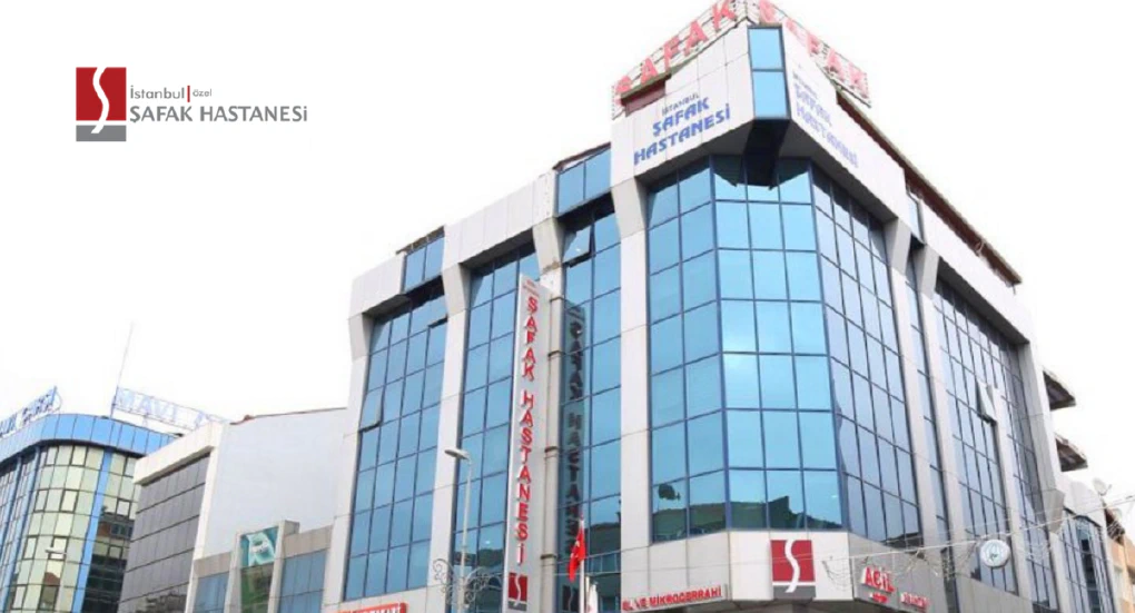
Private İstanbul Şafak Hospital

Our country, which has a strong tourism potential, has become a trend in health tourism with its geographical location, …

1. Pay attention to fluid consumption The calories of the liquid consumed in the summer may not be considered important. …
Contact us before it’s too late for your health.
Don’t delay your health.